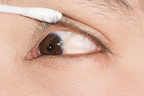
Typically resulting from years of sun exposure, pterygia can be seen as vascular, winged-shaped growths that extend from the white part of the eye (usually the nasal side) towards the colored part.
A pterygium represents the eye’s response to the sun and is often seen in patients who have spent a lot of time outdoors (farmers and surfers, for example).
Symptoms of a pterygium can range from minor irritation and redness to a nagging foreign body sensation or even decreased and distorted vision. Pterygia can be cosmetically bothersome as well.
If a patient’s symptoms are relatively mild, simply using artificial tears (lubricant eye drops) may be sufficient treatment. If the symptoms are more bothersome or if the vision is affected or threatened, surgical removal may be indicated.
The procedure for removing a pterygium is rather straightforward, is performed in an outpatient setting (patient goes home the same day), and requires only local anesthesia (patients receive some sedating medication but almost never require being put to sleep). The procedure normally takes less than 20 minutes to perform. Patients can expect to experience some irritation for several days afterward, but significant postoperative pain is uncommon.
The pterygium procedure is usually done by first removing the lesion (it peels off the surface of the eye), smoothing the underlying area (a special diamond bur is often used), and then gluing a small piece of healthy ocular surface tissue (conjunctiva ) onto to the exposed area previously occupied by the pterygium.
In years gone by, these conjunctival grafts required multiple fine sutures (stitches) to hold the fresh tissue in place. The advent of the tissue glues has rendered the suturing mostly unnecessary, which has shortened the recovery period such that most patients can return to work within a couple of days (depending on the type of work and the severity/size of the pterygium, of course).
Patients will have the eye-patched for the first night and then will begin using eye drops and/or ointments after having the eye patch removed and being examined the day after surgery.
As with any procedure, risks exist. Fortunately, the chance of a serious complication with pterygium surgery is quite low. While recurrences cannot always be prevented, the chance of a significant recurrent pterygium is less than 1% in our practice.
Wearing sunglasses and a hat with a visor when outdoors for prolonged periods seems to help decrease the recurrence rate of an excised pterygium and the growth rate of an existing pterygium.
Even for people who have a small, non-bothersome pterygium, it’s advisable to get the eye(s) examined. Occasionally other more serious conditions can mimic a pterygium – so pleased get checked.
Typically resulting from years of sun exposure, pterygia can be seen as vascular, winged-shaped growths that extend from the white part of the eye (usually the nasal side) towards the colored part.
A pterygium represents the eye’s response to the sun and is often seen in patients who have spent a lot of time outdoors (farmers and surfers, for example). Symptoms of a pterygium can range from minor irritation and redness to a nagging foreign body sensation or even decreased and distorted vision. Pterygia can be cosmetically bothersome as well.
If a patient’s symptoms are relatively mild, simply using artificial tears (lubricant eye drops) may be sufficient treatment. If the symptoms are more bothersome or if the vision is affected or threatened, surgical removal may be indicated. The procedure for removing a pterygium is rather straightforward, is performed in an outpatient setting (patient goes home the same day), and requires only local anesthesia (patients receive some sedating medication but almost never require being put to sleep).
The procedure normally takes less than 20 minutes to perform. Patients can expect to experience some irritation for several days afterward, but significant postoperative pain is uncommon. The pterygium procedure is usually done by first removing the lesion (it peels off the surface of the eye), smoothing the underlying area (a special diamond bur is often used), and then gluing a small piece of healthy ocular surface tissue (conjunctiva ) onto to the exposed area previously occupied by the pterygium.
In years gone by, these conjunctival grafts required multiple fine sutures (stitches) to hold the fresh tissue in place. The advent of the tissue glues has rendered the suturing mostly unnecessary, which has shortened the recovery period such that most patients can return to work within a couple of days (depending on the type of work and the severity/size of the pterygium, of course). Patients will have the eye-patched for the first night and then will begin using eye drops and/or ointments after having the eye patch removed and being examined the day after surgery.
As with any procedure, risks exist. Fortunately, the chance of a serious complication with pterygium surgery is quite low. While recurrences cannot always be prevented, the chance of a significant recurrent pterygium is less than 1% in our practice. Wearing sunglasses and a hat with a visor when outdoors for prolonged periods seems to help decrease the recurrence rate of an excised pterygium and the growth rate of an existing pterygium.
Even for people who have a small, non-bothersome pterygium, it’s advisable to get the eye(s) examined. Occasionally other more serious conditions can mimic a pterygium – so pleased get checked.
